English:
Title: American malacological bulletin
Identifier: americanmal6719881990amer (find matches)
Year: 1983 (1980s)
Authors: American Malacological Union
Subjects: Mollusks; Mollusks
Publisher: (Hattiesburg, Miss. ?) : (American Malacological Union)
Contributing Library: Smithsonian Libraries
Digitizing Sponsor: Biodiversity Heritage Library
View Book Page: Book Viewer
About This Book: Catalog Entry
View All Images: All Images From Book
Click here to view book online to see this illustration in context in a browseable online version of this book.
Text Appearing Before Image:
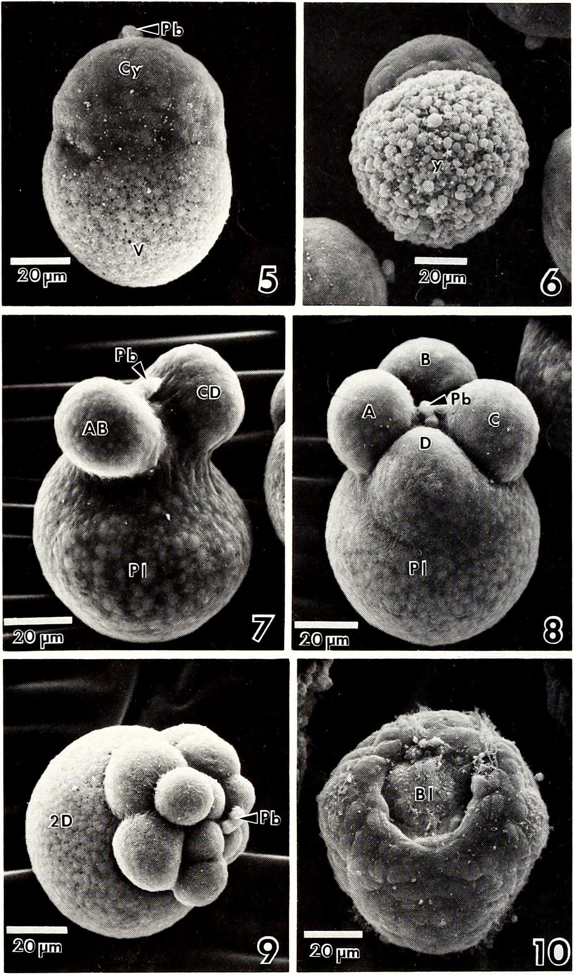
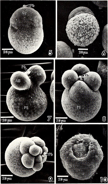
192 AMER. MALAC. BULL. 6(2) (1988) mg; N=40) respectively. The total capsule dry weight varied from 0.92-2.14 mg (x ± S.E.= 1.60 ± 0.06 mg; N=40). Cap- sule wall and content dry weight comprised 65.6 ± 2.3 and 34.4 ± 2.3 (x ± S.E.) percent, respectively, of the total cap- sule dry weight. Capsule wall dry weight varied directly with capsule length: dry weight (mg)= -2.54 + (3.79 X length in cm) (r2=0.673; N = 40; P< 0.001). A significant linear regres- sion of total capsule dry weight on length also existed and is given as dry weight (mg)= -1.48 + (3.28 X length in cm) (r2=0.468; N=40; P0.05). Each capsule contained 3246 ± 21 (x ± S.E.; N = 10) embryos embedded in a viscous, albuminous fluid. Capsules, when deposited, were a milky white color, which during development turned light tan and finally dark brown just prior to hatching. Only three capsules deposited in the laboratory developed the dark purple color, characteristic of dead or stressed embryos (St. Amant, 1938; D'Asaro, 1966; Spight, 1977; Pechenik, 1982; Butler, 1954, 1985). Examina- tion of these capsules revealed that all embryos were dead. DEVELOPMENTAL RATE AND STAGES Development of Thais haemastoma canaliculata was synchronous within a particular capsule throughout the en- tire period of encapsulation and required 12-13 days to hatch- ing at 25°/00S and 25°C (Table 1). Unfertilized eggs were spherical and approximately 65-70 fivr\ in diameter; however, as reported previously (St. Amant, 1938; DAsaro, 1966), the majority of the yolk (deutoplasm) was concentrated in one pole (vegetal) with other cytoplasmic constituents being concen- trated at the opposite (animal) pole. First and second polar body formation was complete within 2.5 hours after deposi- tion of the capsule. By the second polar body stage (Fig. 5), the fertilized egg had elongated and the animal and vegetal areas were easily distinguished. The round yolk granules in the vegetal area were visible in live and preserved (Figs. 5, 6) zygotes. Early cleavage was restricted to the animal pole of the embryo. The first cleavage, producing the AB and CD blastomeres (Fig. 7) occurred 5-6 hours after deposition (Table 1). The second cleavage (Fig. 8) occurred within 2-4 hours after the first cleavage. As DAsaro (1966) showed for T, Table 1. Developmental rate of Thais haemastoma canaliculata at 25°/00S and 25-26°C. Developmental Event Time Fertilized egg with 2 polar bodies 2.5 hours First cleavage 5-6 hours Second cleavage 8-9 hours 16 cell stage 17-19 hours Stereoblastula 28 hours Early gastrula 3.5-4 days Stomodael invagination, cephalic expansion & shell gland formation 5 days Trochophore 5.5-6 days Early veliger 7 days Hatching 13 days haemastoma floridana, we found that the D blastomere possessed a large polar lobe (Fig. 8). Within 17-19 hours after capsule deposition, the 18 cell stage was complete. By that time, the polar lobe had been resorbed, and the large 2D macromere was seen (Fig. 9). A stereoblastula containing a narrow segmentation cavity, as reported by St. Amant (1938), formed approximate- ly 9-11 hours after polar lobe resorption (Table 1). Gastrula- tion by epiboly and archenteron formation (Fig. 10) was
Text Appearing After Image:
Fig. 5. SEM of fertilized egg after second polar body formation (Cy, cytoplasmic (animal) pole; Pb, polar bodies; V, vegetal yolk-containing pole). Fig. 6. SEM of vegetal view of ruptured polar lobe, illustrating dense yolk mass (y, yolk mass). Fig. 7. SEM showing first cleavage of the ovum, resulting in formation of AB and CD cells (Pb, polar bodies; PI, polar lobe). Fig. 8. SEM of four-cell stage showing com- pletion of A, B, C, and D cells with polar lobe (PI) and polar bodies (Pb) still evident. Fig. 9. SEM of 2D cell, after polar lobe resorption (Pb, polar bodies). Fig. 10. SEM of gastrula stage, illustrating the blastopore (Bl).
Note About Images
Please note that these images are extracted from scanned page images that may have been digitally enhanced for readability - coloration and appearance of these illustrations may not perfectly resemble the original work. 
_(18157588551).jpg)

_(18157588551).jpg&action=edit&withJS=MediaWiki:Catcheck.js)

_(18157588551).jpg)