
Bestand:Trichinella spiralis (07).tif

Oorspronkelijk bestand (1.481 × 2.585 pixels, bestandsgrootte: 2,58 MB, MIME-type: image/tiff)
| Dit is een bestand van Wikimedia Commons. Onderstaande beschrijving komt van de beschrijving van het bestand daar. |
Beschrijving
| Beschrijving |
On the anatomy, decay and development of the trichina spiralis. "From the Transactions of the Pathological Society for 1854." DESCRIPTION OF PLATE XIII. The figures illustrate Dr. Bristowe’s (and Mr. Rainey’s) Observations on the Anatomy of the Trichina Spiralis. Page 274. Fig, 1. Trichina spiralis. a a a integument; b b b. muscular layer; c mouth; d, anus; e. oesophagous; f f. alimentary canal; g. funnel shaped portion, with pyriform bodies at its base; h. tube connected with reproductive process; i. yellow deposit in it; j j. space between mus- cular layer and parts internal to it. It terminates above in minute points. Fig. 2. The same less magnified, intended to show better the relative size and situation of parts, The letters indicate the same structures as in the preceding. Fig. 3. The same with its surface in focus, to show one of the longitudinal muscular bands, Fig. 4. Fragment of the anterior part of the worm, with the alimentary canal protruding, and apparently exhibiting a central tube. Fig. 5. Fragment of posterior part of worm. The generative tube, h protrudes, and its cellular structure is exhibited. A portion of the alimentary canal is likewise exposed. At this part it is contracted, and consists apparently of a structureless membrane studded with refractive globules. Fig. 6. Generative tube acted on by acetic acid. The cells are heaped together, and the basement membrane is seen. Fig. 7. Integuments of the lower part of the worm from which the entire contents have been separated by endosmose. The crenate appearance lost, and the prolongation into the anus is visible. Fig. 8, Appearance frequently presented by fat formed either free among the muscles, or connected with Trichina cysts. |
| Datum | |
| Bron | https://archive.org/stream/b21476809#page/n5/mode/2up |
| Auteur | Rainey, G. (George), 1801-1884, Bristowe, John Syer, 1827-1895. Pathological Society of Great Britain and Ireland. University of Glasgow. Library. Publisher: [London] : [Printed by J.W. Roche] |
|
Dit werk bevindt zich in het publiek domein in landen en gebieden waar de auteursrechttermijn het leven van de auteur plus 70 jaar of minder is.
| |
| Van dit bestand is vastgesteld dat er geen bekende auteursrechtaanspraken op rusten, alle aanverwante en naburige rechten daarbij inbegrepen. | |
Bestandsgeschiedenis
Klik op een datum/tijd om het bestand te zien zoals het destijds was.
| Datum/tijd | Miniatuur | Afmetingen | Gebruiker | Opmerking | |
|---|---|---|---|---|---|
| huidige versie | 27 sep 2019 16:15 | 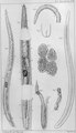 | 1.481 × 2.585 (2,58 MB) | Rasbak | grey |
| 26 sep 2019 18:53 |  | 1.481 × 2.585 (7,19 MB) | Rasbak | improved color | |
| 26 sep 2019 08:39 | 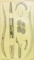 | 1.481 × 2.585 (6,81 MB) | Rasbak | {{Information |Description=On the anatomy, decay and development of the trichina spiralis. "From the Transactions of the Pathological Society for 1854." DESCRIPTION OF PLATE XIII. The figures illustrate Dr. Bristowe’s (and Mr. Rainey’s) Observations on the Anatomy of the Trichina Spiralis. Page 274. Fig, 1. Trichina spiralis. a a a integument; b b b. muscular layer; c mouth; d, anus; e. oesophagous; f f. alimentary canal; g. funnel shaped portion, with pyriform bodies at its base; h. tube c... |
Bestandsgebruik
Dit bestand wordt op de volgende pagina gebruikt: